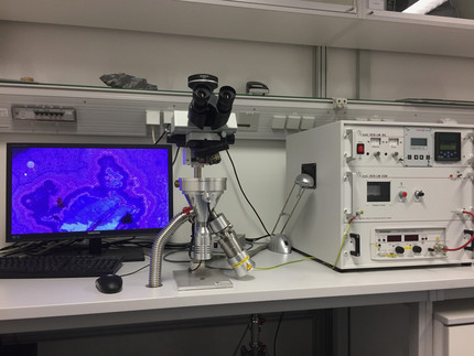
Cathodoluminescence microscopy (CL)
Cathodoluminescence microscopy is a technique to visualise in minerals and materials heterogeneities and internal structures that are invisible using pure optical methods (reflected and transmitted light microscopy).
An electron source (electron gun with W-filament) generates an electron beam that is directed onto the solid-state sample (polished and carbon-coated thin section) and excites it. Subsequent transition from excited to normal state causes the emission of light (luminescence) that can be observed in true colour and intensity using a polarising microscope. Internal structures of minerals become visible and are indicative of growth zoning, trace element zoning and structures allowing processes such as mineral growth, resolution and alteration to be reconstructed. Furthermore, it helps to rapidly distinguish optical similar minerals, like calcite-dolomite or monazite-xenotime.
Instruments
Olympus BXFM-F polarising microscope, equipped with:
- Lumic hot-cathode electron source
- Olympus XC10 digital colour camera, very high sensitivity

