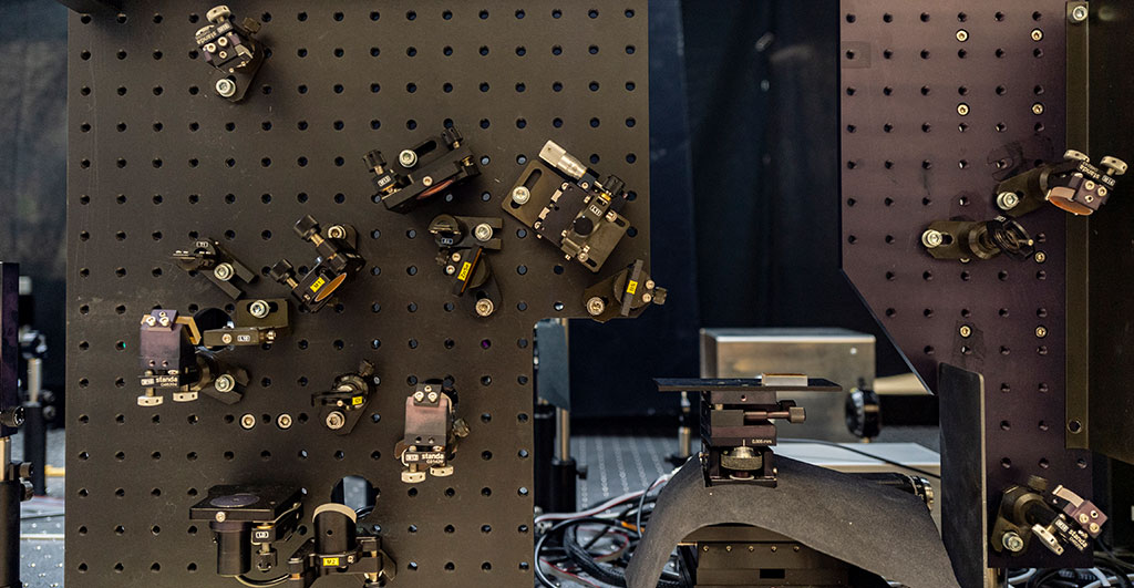
“I like complicated experiments,” says the junior professor of physical chemistry. Standing in her laser lab, it quickly becomes clear what she means. Half of the room is taken up by a seven-meter-long table with wired boxes bolted to it. A look inside the open boxes reveals lots of round and angular metal elements, prisms, mirrors, lenses, and crystals. The layperson quickly loses track and is not able to recognize the pattern in which they are arranged. But what can be seen here follows a sophisticated plan. Every little screw is in the right place, every mirror and every lens is aligned with high precision.
Whether everything is in place and every angle is correct becomes apparent when an ultrashort laser beam is fed into the system. The pulsed light beam with a wavelength of 800 nanometers is visible as reddish light. It is split and bundled with the help of lenses, redirected via mirrors, refracted in the inserted crystals, and it changes its wavelength. When the light has been bent, refracted, bundled, and manipulated multiple times, mid-infrared light is produced at the end of the assembled laser system, which Müller-Werkmeister and her research team can use to observe the movement of molecules.
Bonds break and form again
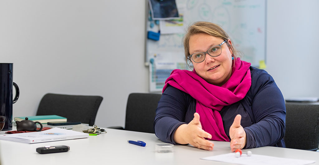
Müller-Werkmeister holds the model of a water molecule in her hands and shows what she means by this: A red sphere represents the oxygen atom, two white spheres are the hydrogen molecules bound to it. She briefly pulls on one of the white spheres, which detaches from the model with a “plop”. “That’s a bond break,” she explains. In chemistry, that’s nothing special. Molecules and atoms are constantly in motion, breaking bonds and forming new ones, changing their structures, releasing or absorbing energy. “Chemical processes like these are written down on paper as mechanisms in organic or inorganic chemistry. But until now, we have not been able to observe them, for example in the synthesis of pharmacologically active substances or new materials,” the physical chemist explains.
The researcher wanted to change just that. “How can I study biomolecules on the fundamental time scale of chemistry?,” she asked herself. To do that, she needs light of a very specific quality – with the right wavelength, intensity, and pulse rate. “Technically, it is very difficult to produce this ultra-fast infrared light,” Müller-Werkmeister explains. Over four years ago, the scientist started to retool the laser laboratory in such a way that completely new insights into biomolecules, such as proteins, sugars, and enzymes, have become possible. The first measurements were made a year ago using a method called ultrafast 2D infrared spectroscopy (2D IR spectroscopy). Only about ten research groups in Germany are working on this method, and only three groups are working on biomolecules.
Here, “ultrafast” means: damned fast. Light travels just 0.33 micrometers in a femtosecond; a femtosecond is the millionth part of a billionth of a second. “From a chemical perspective, this is a very interesting time scale, but one that is not at all easy to study,” Müller-Werkmeister explains.
Vibrations in the molecule soup
She has now achieved this feat with the laser system in her lab and ultrafast 2D IR spectroscopy, which makes the structural changes – the dance of molecules – visible. To do this, the light beam hits a “molecular soup” of millions of molecules, as she calls it, and induces vibrations in the compounds. These vibrations provide signals that are visualized as a spectrogram. The method looks at very specific groups of atoms, known as functional groups, that are important to the molecules’ reaction behavior and properties. “It’s as if we’re looking at an entire lecture hall full of students, but we’re specifically only looking at their hairstyles,” Müller-Werkmeister says, describing the principle.
It becomes particularly interesting for her when the “hairstyles” change after a certain time: What do the vibrations of the molecules look like after a set time, and what does that reveal about their properties and possible interactions with other substances? “Molecular processes start on very short time scales, after one second you already have a completely different molecule,” she explains. “We are interested in what exactly happens at the molecular level during this time.” These intermediate steps, after all, are often still unknown. Therefore, she is planning a series of measurements for the future that can cover entire reaction sequences. For example, what happens during the chemical synthesis of a substance after 100 femtoseconds, a nanosecond or a millisecond, and what exactly changes in the molecule?
The fingerprint of microplastic
The establishment of the laboratory for 2D IR spectroscopy in Potsdam took years of work. It is a process that has not yet been completed and that is constantly being refined. For the research team led by Müller-Werkmeister, the effort is worthwhile. This is because the method offers a kind of “toolbox” that can be applied to all kinds of research questions. The overall goal is to better understand chemical and biochemical reactions and the associated structural changes. Right now, for example, the team is optimizing IR spectroscopy for very specific functional groups that often exist in pharmacologically active molecules. “Phosphate groups, sulfate groups, or fluorinated carbons are highly interesting, active in many biomolecules, and have only been studied by three other research groups worldwide,” Müller-Werkmeister emphasizes. If more were known about the molecule behavior, this could also provide new insights into how exactly they are effective against diseases or influence cell functions.
In addition to sugar molecules and proteins, the researchers also focus on microplastics because each type of plastic has an individual fingerprint in IR spectra. “The big challenge is to first isolate the plastic particles from environmental samples – for example, from arable land,” Müller-Werkmeister explains. Her research group succeeded with a combination of different methods: The coarser plastic particles were first separated from the soil particles through sieving. The remaining particularly small particles could then be electrostatically charged and pulled out of the soil as with a magnet. Also due to the different density of plastic and soil particles, both could be separated from each other in one process. Up to 25,000 samples containing plastic particles smaller than 15 micrometers can then be measured on a specially developed silicon chip. In this size range, the particles can be hazardous to organisms and the environment, and no method exists yet that can analyze large numbers of samples in a short time. From the specific features of the spectrum, the researchers can identify exactly what type of plastic is included and also estimate the approximate age of the samples. The research team is currently refining the methods in order to be able to analyze large quantities of soil samples in a short time.
Whether it's biomolecules, microplastics, or metallic nanoparticles, Müller-Werkmeister, who is involved in the UniSysCat Cluster of Excellence with her project researching the green chemistry of the future, has many plans for what else she would like to investigate in her laser laboratory. In the near future, she plans to combine results from ultrafast spectroscopy and X-ray crystallography. The latter provides extremely high-resolution information about molecules down to the atomic level. For the fast X-ray experiments, she is collaborating with researchers at the German Electron Synchrotron (DESY) in Hamburg to gain even more detailed insights into the structure and reaction mechanisms of biomolecules. With this research project, too, she is breaking new ground. But that doesn’t deter the junior professor. After all, she’s not afraid of complicated experiments.
The Lab
The 2D IR spectroscopy laboratory of the group is equipped with high-precision and state-of-the-art measurement technology, including a laser system with a two-stage regenerative amplifier, optical parametric amplifiers for multi-stage conversion to mid-infrared and/or variably tunable visible light, difference-frequency generation for mid-infrared, a 2D IR spectrometer with a pulse shaper, and an MCT detector.
The setup is funded by the tenure track program of the University of Potsdam and the State of Brandenburg and by the funding guidelines EFRE InfraFEI and EFRE StaF of the European Union.
The Researcher
Prof. Dr. Henrike Müller-Werkmeister studied biochemistry and physics at Goethe University Frankfurt. Since 2017, she has been Junior Professor and head of a group in physical chemistry working on ultrafast structural dynamics.
Mail: henrike.mueller-werkmeisteruuni-potsdampde
This text was published in the university magazine Portal Wissen - Two 2022 „Humans“ (PDF).