


Single particle cryo EM
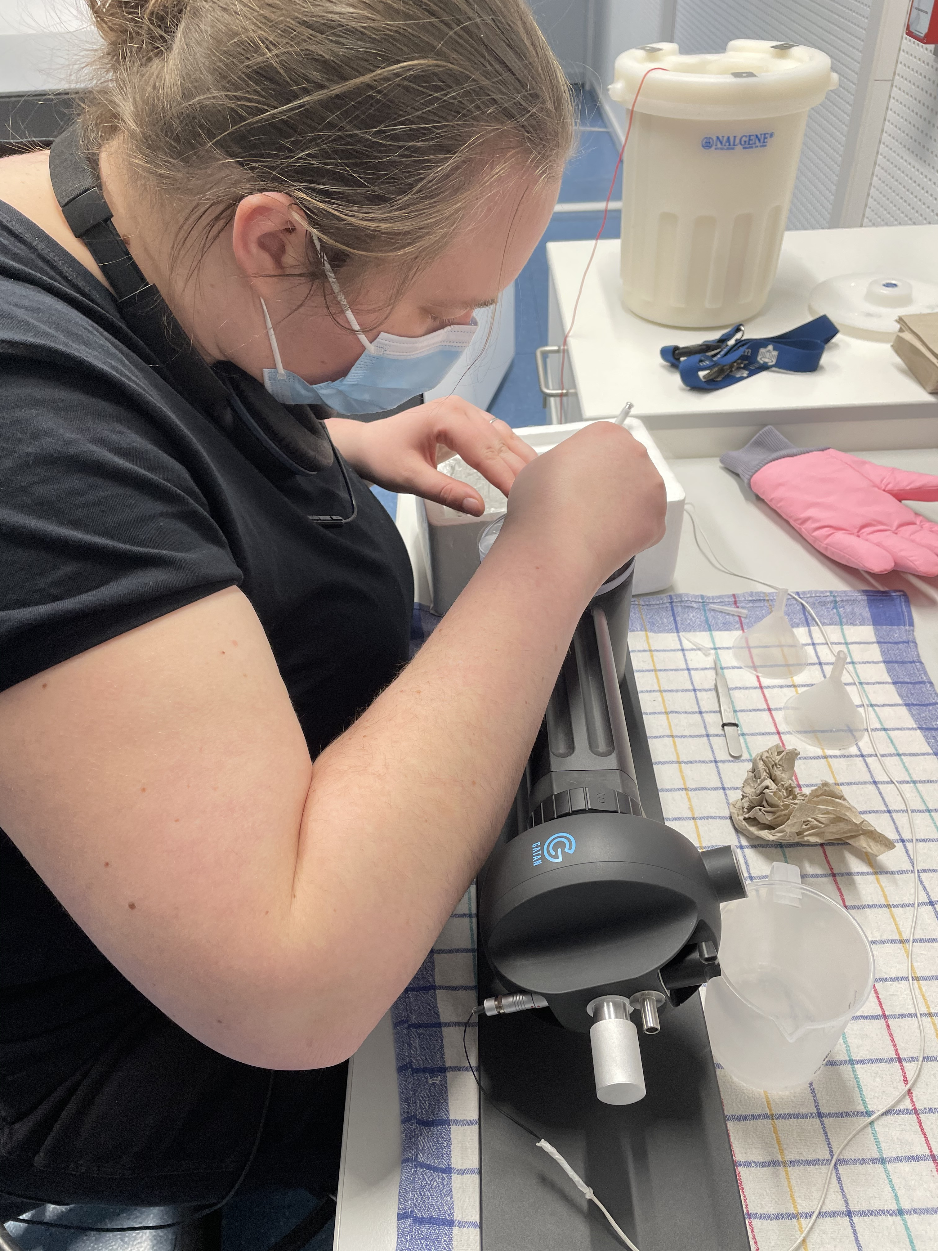
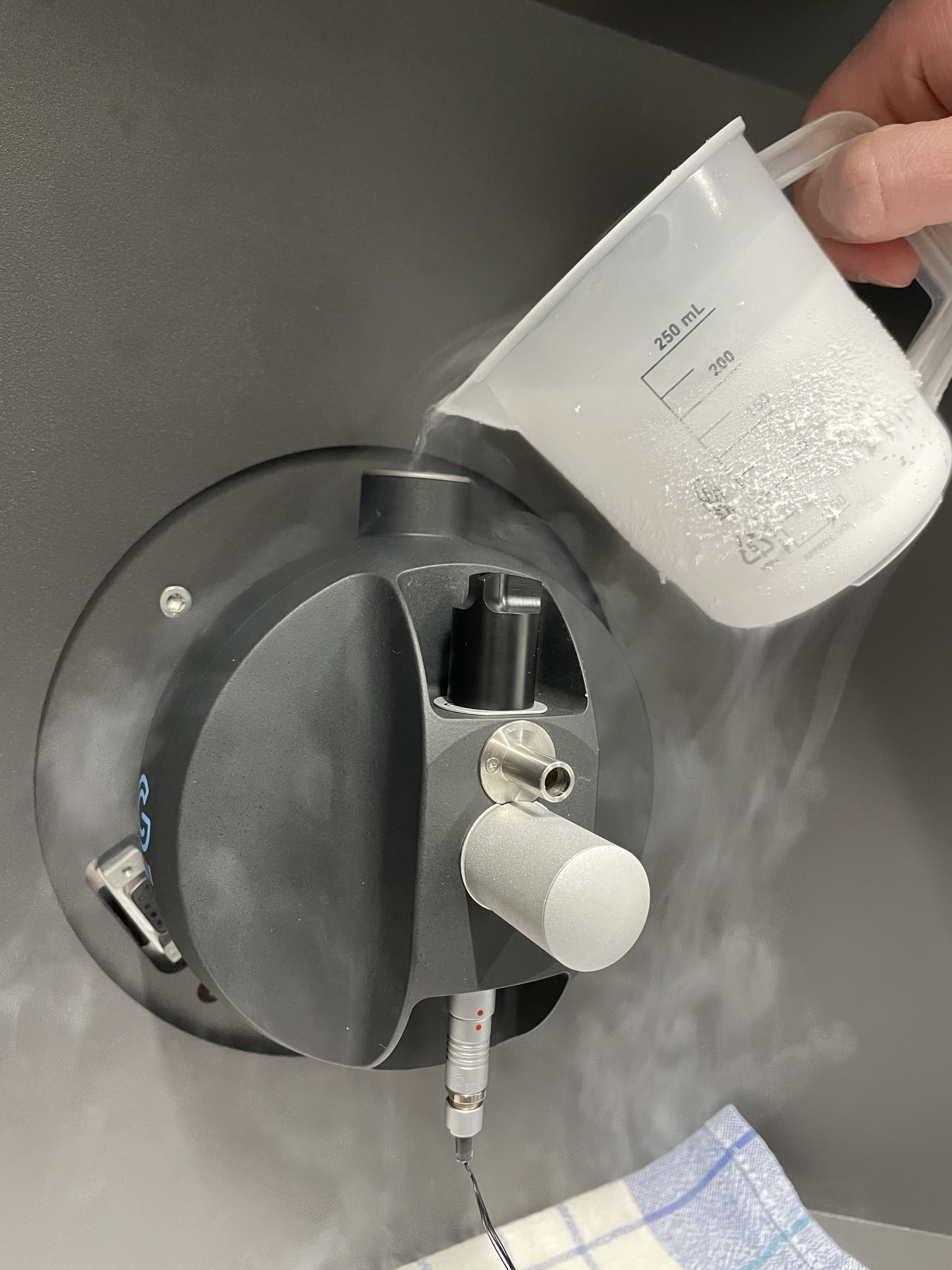
Cryogenic electron microscopy (cryo EM) allows structural investigation of macromolecules at atomic resolution, which is necessary to explore the very architecture of life. In biological sciences it recently has attracted wide attention as an alternative to x-ray crystallography or NMR spectroscopy for protein structure determination (1, 2), because it requires only a small amount of sample and can work for heterogenous or flexible complex mixtures. In brief, the aqueous protein sample is plunge frozen in liquid ethane, ideally by adopting random orientations in the ice layer. Thousands of particle images are digitally recorded in the electron microscope and the three-dimensional (3D) structure of the protein complex is calculated using powerful CPU or GPU image processing analyses. Recently, the technique attained the ability to locate individual atoms within a protein (3).
We use cryo electron microscopy (EM) to investigate the high-resolution 3D structure of macromolecular complexes, such as AAA+ proteins, redox protein complexes, proteasomes, or small membrane complexes. We are particularly interested in the dynamic movements of macromolecular complexes during their biological activity and aim to obtain molecular movies for these nano machines.
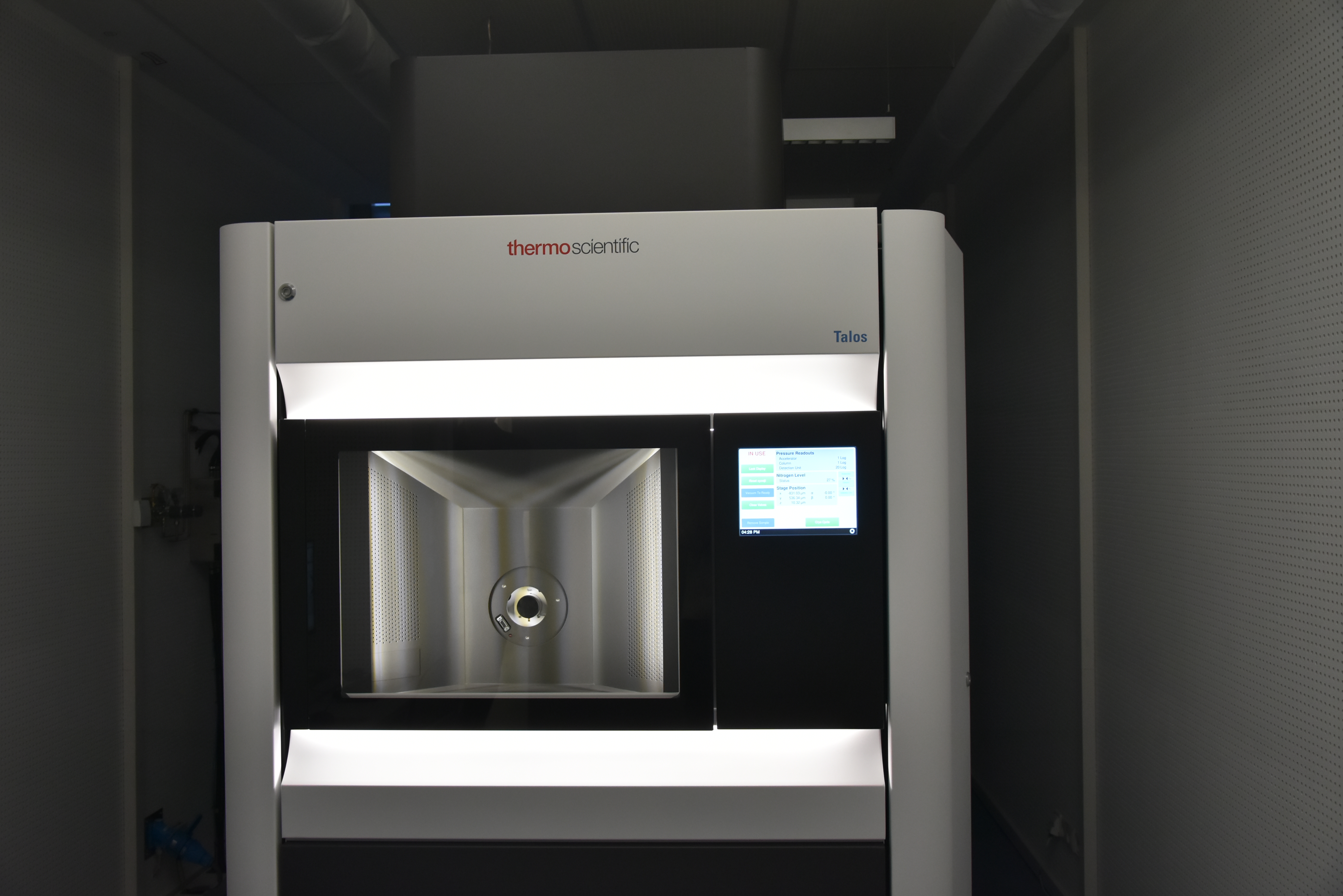
The University of Potsdam owns a 200 keV Talos F200C G2 (Thermo Fisher) electron microscope equipped with a Falcon III direct electron detector and Ceta M16 camera for cryo EM sample screening and data collection. Two cryo EM holders for single grid (ElsaTM, Gatan) or multi-grid (Simple Origin) data collection for up to 9 hours can be used. Grid preparation equipment such as Zepto (Diener) and Yocto (Diener) glow discharge units and Leica EM GP 2 (Leica) grid plunger are available at the facility. The faculty of science at the University of Potsdam maintains a compute cluster consisting of 3 CPU based nodes and one GPU based node that is at disposal of the Wendler group. Furthermore, several smaller servers with a total of 12 high-end GPU cards and 150 TB of storage fileservers are owned by the Wendler lab. We use most of the established cryo EM software packages available for image processing and molecular modelling on our Linux clusters or 3D Graphics machines.
Microscope booking conditions and user fees are regulated by the terms of use. The facility is only available to internal users (members of the University of Potsdam). Please contact Petra Wendler if you are interested in using the EM facilty.

Protein Purification
We purify macromolecular complexes from E. coli, S. cerevisiae or insect cells either after overexpression of heterologous proteins or by tagging of endogenous proteins. Thus, we use all standard molecular biological, biophysical and protein biochemical techniques such as cloning, PCR, Immunoprecipitation, size exclusion chromatography, density gradients, electrophoresis, Western blot, surface plasmon resonance spectroscopy and fluorescence microscopy. For functional analyses in yeast, we manipulate their genome so that complementation, deletions or mutations can be analysed.
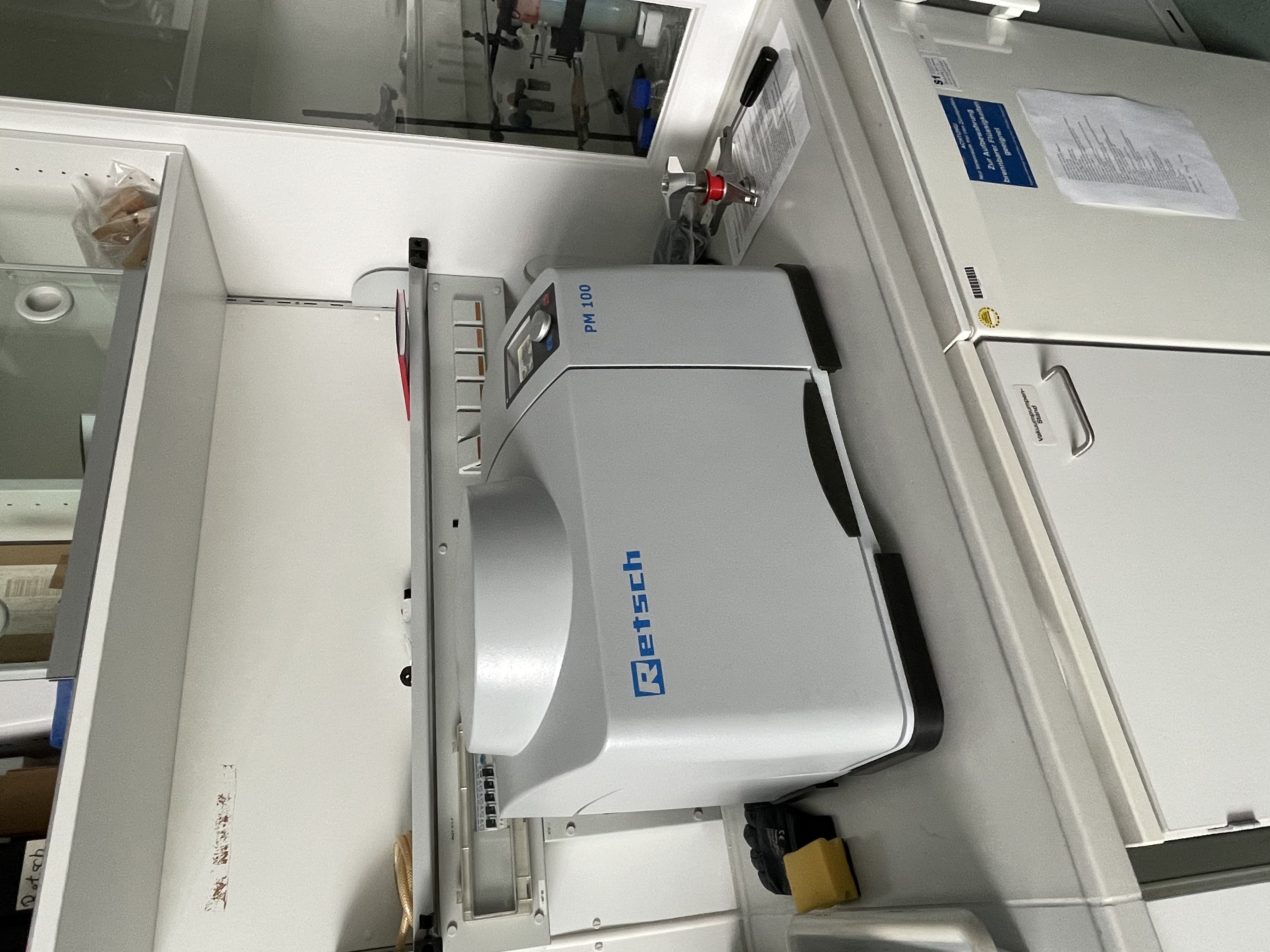
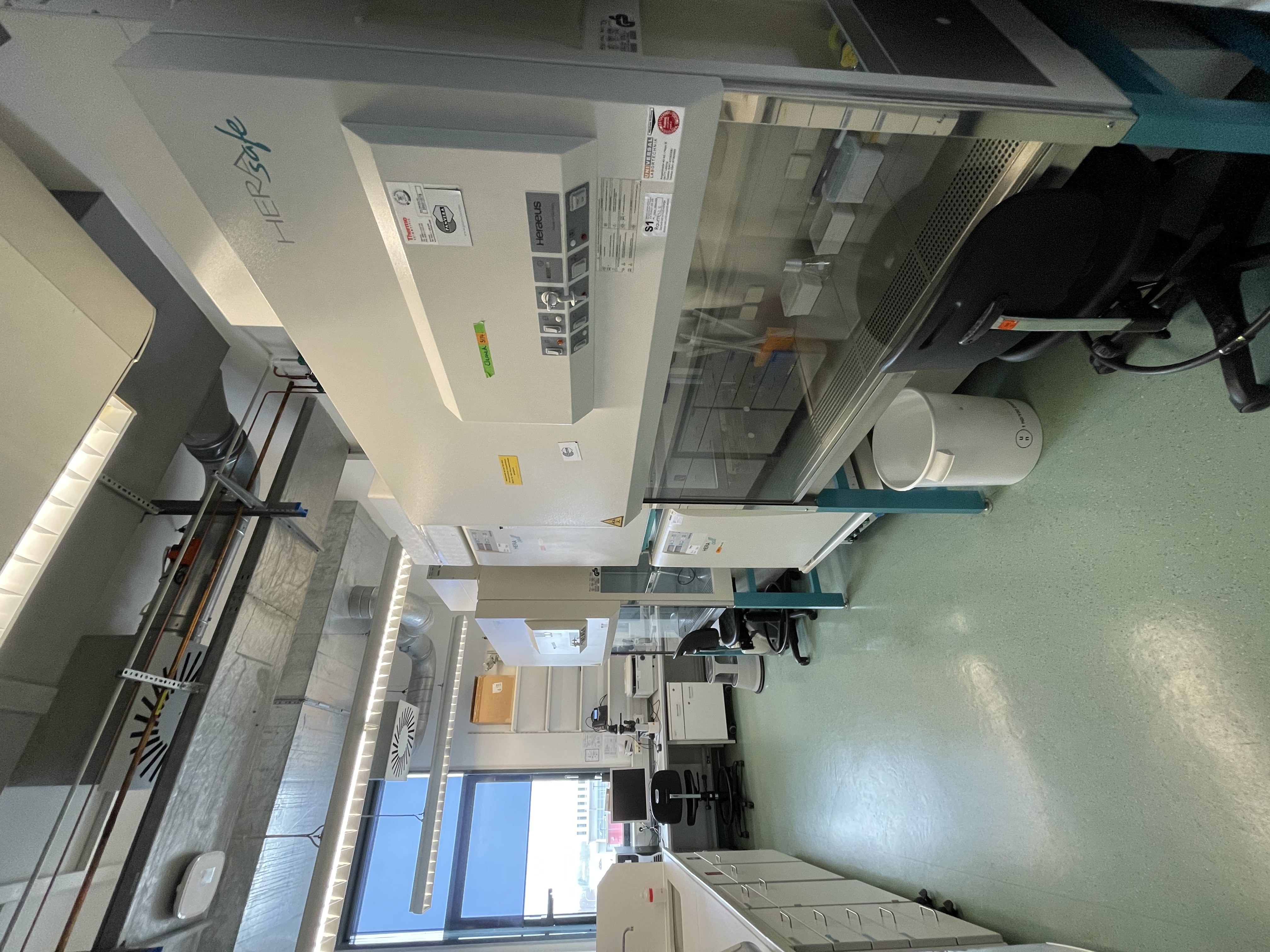
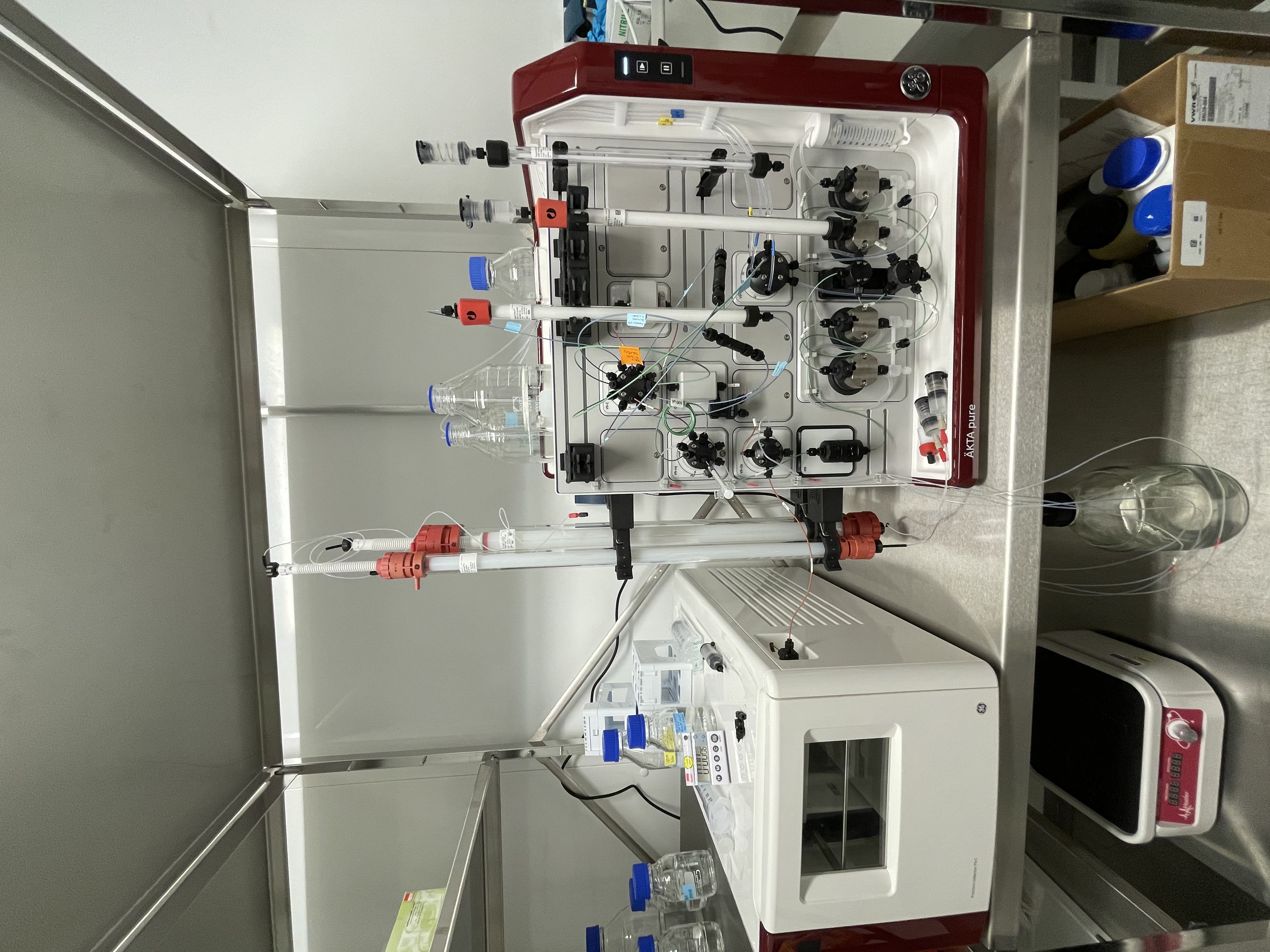
Our labs are equipped with several Innova Shakers for large scale cultivation of bacterial or yeast cells. The cell culture lab hosts different Eppendorf incubators and 2 Herasafe Celan benches. Protein extraction can be performed using a MM400 mixer mill (Retsch), a PM100 mill (Retsch), or an Aminco French Press. The lab is also equipped with an OPTIMA L-100 XP (Beckman) ultracentrifuge and an ÄKTA Pure FLPC (G&E) system. All equipment necessary for standard cloning techniques and protein detection is available in the lab.
Further Techniques
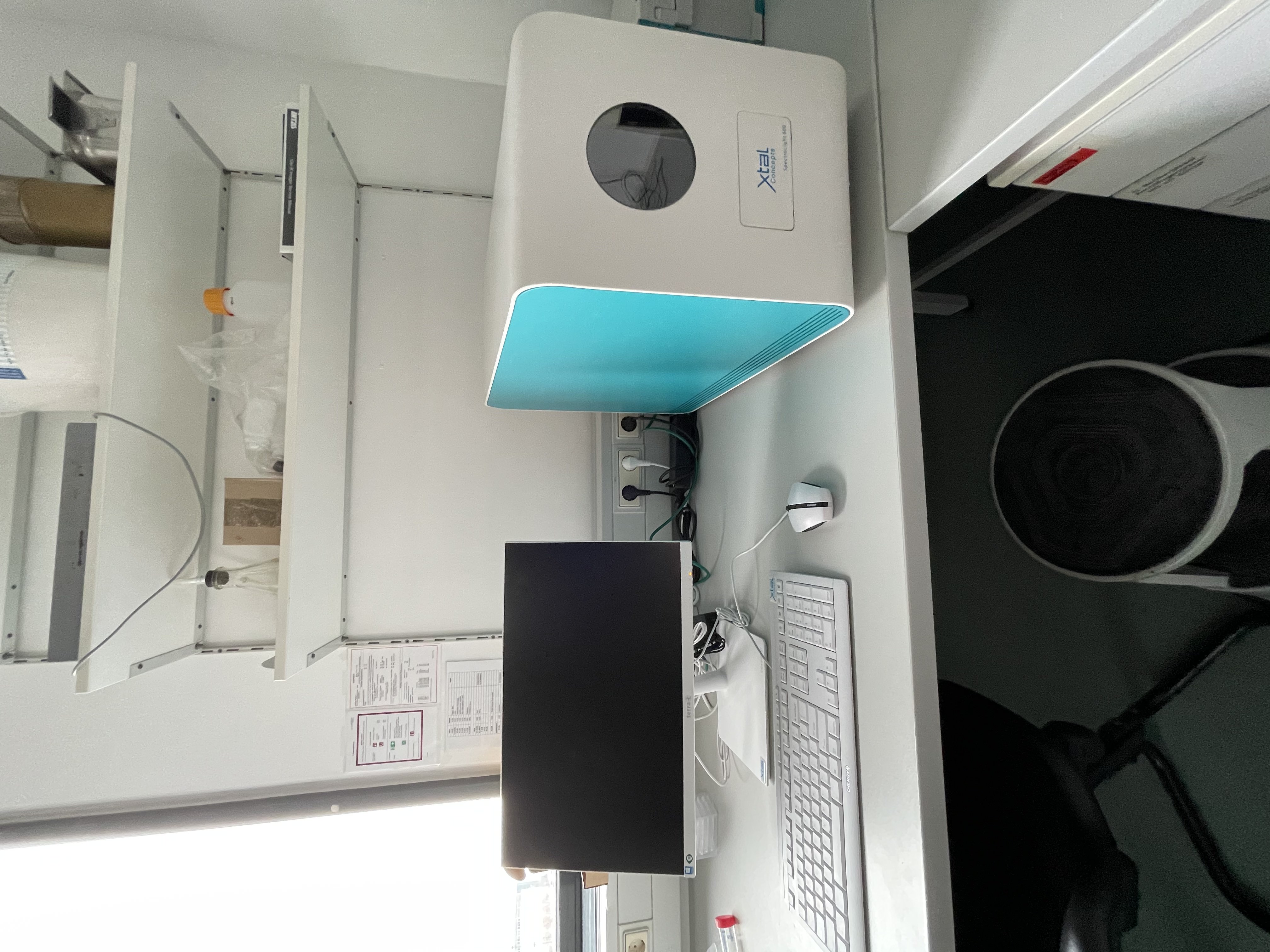
In 2020, we obtained a Spectrolight 610 (x-tal concepts) in-situ dynamic light scattering instrument that we use to assess complex homogeneity and assembly state, ideal storage buffer composition, nanodisc/protein assembly, long time protein stability, protein crystallisation conditions and many more protein properties.
1: Cressey D, Callaway E (October 2017). "Cryo-electron microscopy wins chemistry Nobel". Nature. 550 (7675): 167. doi:10.1038/nature.2017.22738
2: Kühlbrandt W (2014). “The Resolution Revolution”. Science 343 (6178): 1443-1444. DOI: 10.1126/science.1251652
3. Herzig M A (October 2020). „Cryo-electron microscopy reaches atomic resolution“. Nature. 587, 39-40. DOI: https://doi.org/10.1038/d41586-020-02924-y