
“The idea of the project SMART-DNA is to use DNA material to produce nanostructures,” says Ilko Bald, leader of the working group Hybrid Nanostructures at the Institute of Chemistry. DNA is a stable biomolecule that can be produced artificially and is easy to work with. Since it is intended as a carrier of genetic information for information processing, it can be used as a programmable material. “We have DNA artificially produced by a company. The three-dimensional shape of the nanostructure is programmed into the material,” he summarizes. “We can then use this tool to perform measurements on molecules.”
Focusing on molecules
To obtain information about chemical reactions of molecules in a solution, the researchers use Raman spectroscopy. Here, the investigated substance is irradiated with laser light of a specific wavelength. A small portion of the light that is scattered back by the substance changes its wavelength due to interaction with the sample. The intensity of the scattered light results in an individual Raman spectrum for each chemical substance.
This method, however, is not very sensitive, which is why a very high molecular concentration is required for the analyses. Yet, with the help of nanoparticles, usually of gold or silver, that are docked onto the DNA structure, Raman spectroscopy becomes extremely sensitive. “The nanoparticles amplify the light scattering and act like an antenna,” Bald explains. “They make it possible to record spectra from the molecules that are situated between two particles.” Whereas the conventional method still requires millions of molecules, surface-enhanced Raman scattering can even record individual molecules. “This is not possible with any other method,” he emphasizes.
Origami with DNA
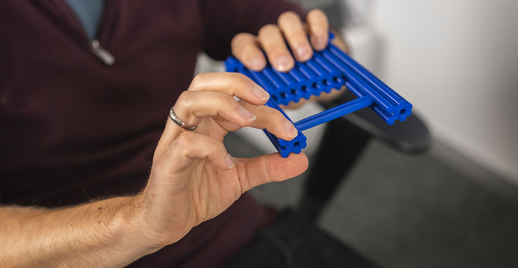
The challenge is to find suitable nanoparticles and to place the molecules exactly where the signal is amplified the most. Prof. Bald shows a DNA model from the 3D printer. “We designed this structure ourselves. Under the DNA bridge, you can insert single molecules exactly between the nanoparticles because that’s where we want to measure Raman scattering.” Using a technique known as DNA origami, a long strand of DNA double helix can be folded several times until the desired nanostructure is achieved. Since there should be as little interfering material as possible around the molecules to be measured, there is a material-free “window”. A stable framework supports the nanoparticles, and a base fixes the structure on the surface.
For the initial Raman studies, Bald and his team had previously worked with a two-dimensional triangular structure, but this had certain disadvantages. That is why a new shape was necessary. The team invested about three years of work in designing and optimizing this structure. “Now we can actually take measurements with it and routinely introduce relatively complex molecules such as proteins into these structures,” he says, pleased.
Sensitive detection methods for medical diagnostics
Measuring individual molecules is of great interest for diagnostic procedures in the biomedical field. “We are currently working on how receptors could be attached to our nanostructures in order to detect biomarkers, for example,” Bald explains one possible application. In low concentrations, biomarkers already form in the body very early, for example when a tumor develops. In addition, the single-molecule technique can be used to understand the process of biochemical reactions step by step. In the future, Prof. Bald would like to make the nanostructures available to other working groups as a tool for a basic research laboratory in order to facilitate collaboration on applied issues.
The Researcher
Prof. Dr. Ilko Bald studied chemistry at Freie Universität Berlin. From 2013, he was Junior Professor for Optical Spectroscopy and Chemical Imaging and since 2019, he has been Professor for Hybrid Nanostructures at the University of Potsdam.
Mail: ilko.balduuni-potsdampde
The Project
“SMART-DNA: Single-Molecule Analytical Raman Tools based on DNA nanostructures has been funded by an ERC-Consolidator Grant” since 2018. The grant is endowed with about 2 million euros for the establishment of a research group of one’s own.
Participants: Universität Potsdam
Funding: European Research Council – ERC
Duration: April 2018–March 2023
Observing nanostructures
Since the size of the examined nanostructures ranges from 10 nanometers (1x10⁻⁸ meters) to 100 nanometers (1x10⁻⁷ meters), they are observed with an atomic force microscope. For this purpose, the surface is scanned with a kind of tiny record needle attached to a leaf spring. With the help of a laser, the bending of the spring can be measured and an image of a surface profile is created.
This text was published in the university magazine Portal Wissen - Eins 2023 „Lernen“ (PDF).