Available methods
Spectroscopy

CD spectroscopy
Structure determination of chiral (bio)molecules and investigation of their interactions with other molecules
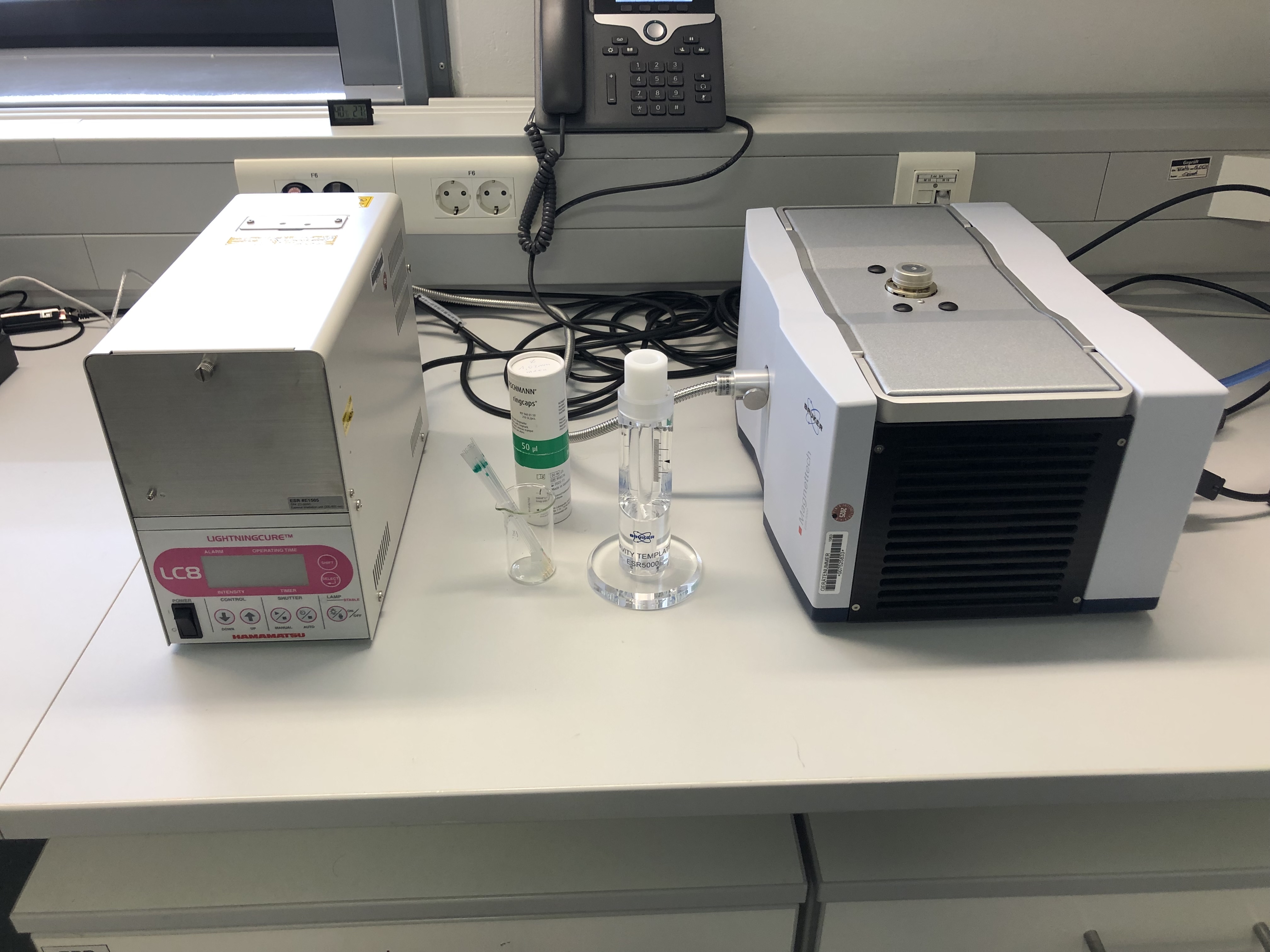
EPR spectroscopy
Electron spin resonance measurements of paramagnetic substances, including under irradiation
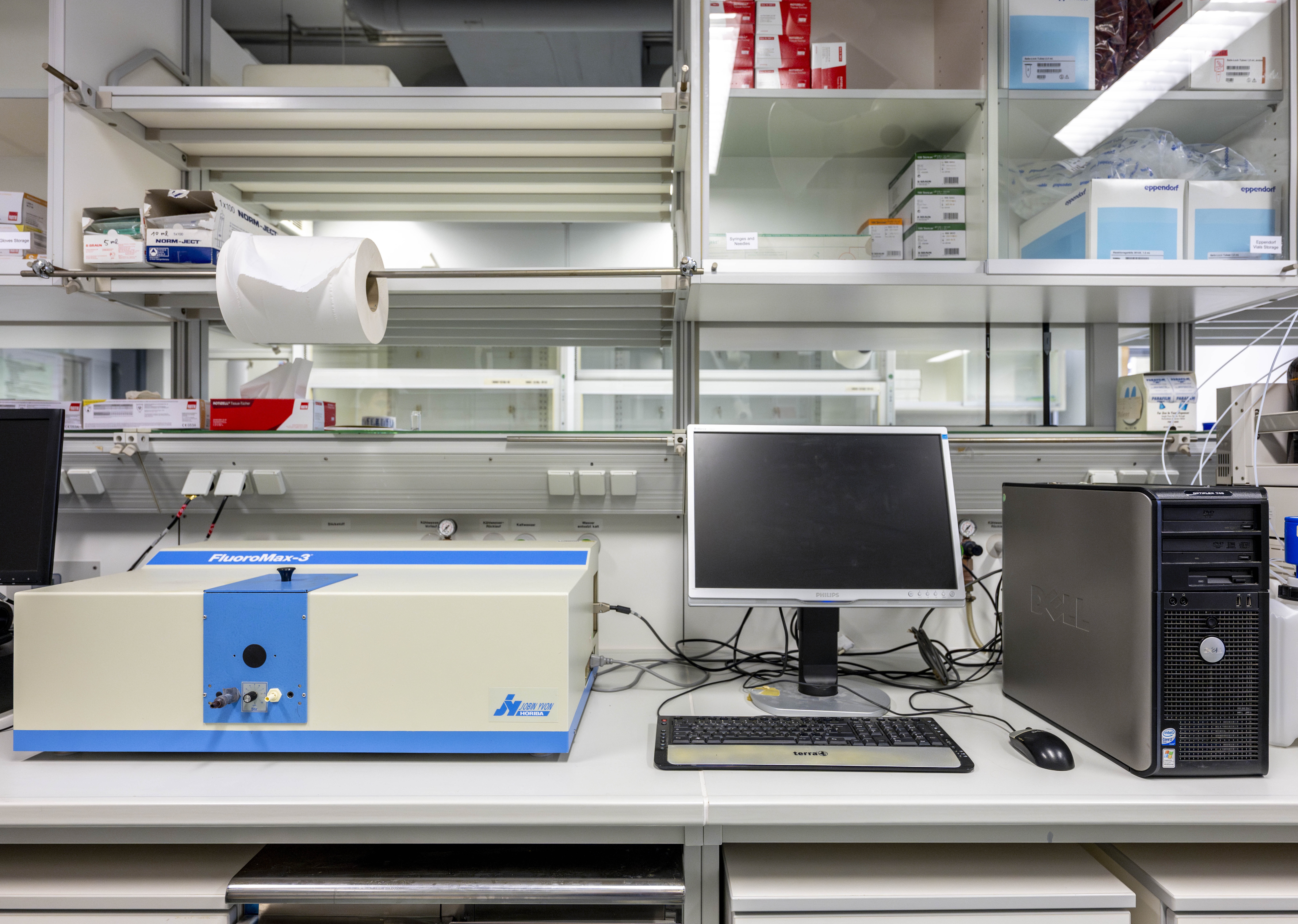
Fluorescence spectroscopy
Fluorimetric measurements
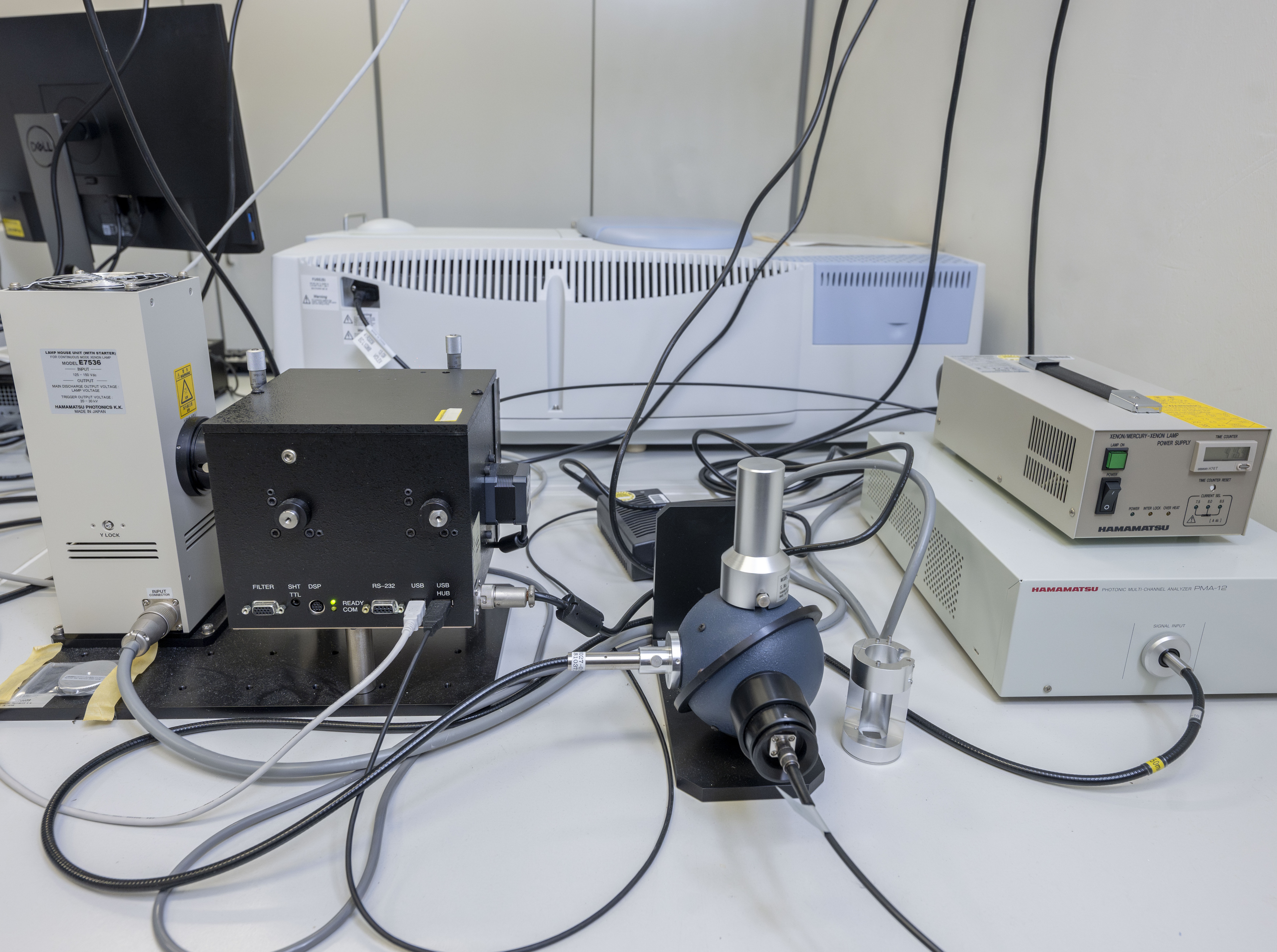
Fluorescence quantum yield
Determination of the quantum yield in the range 300-950 nm
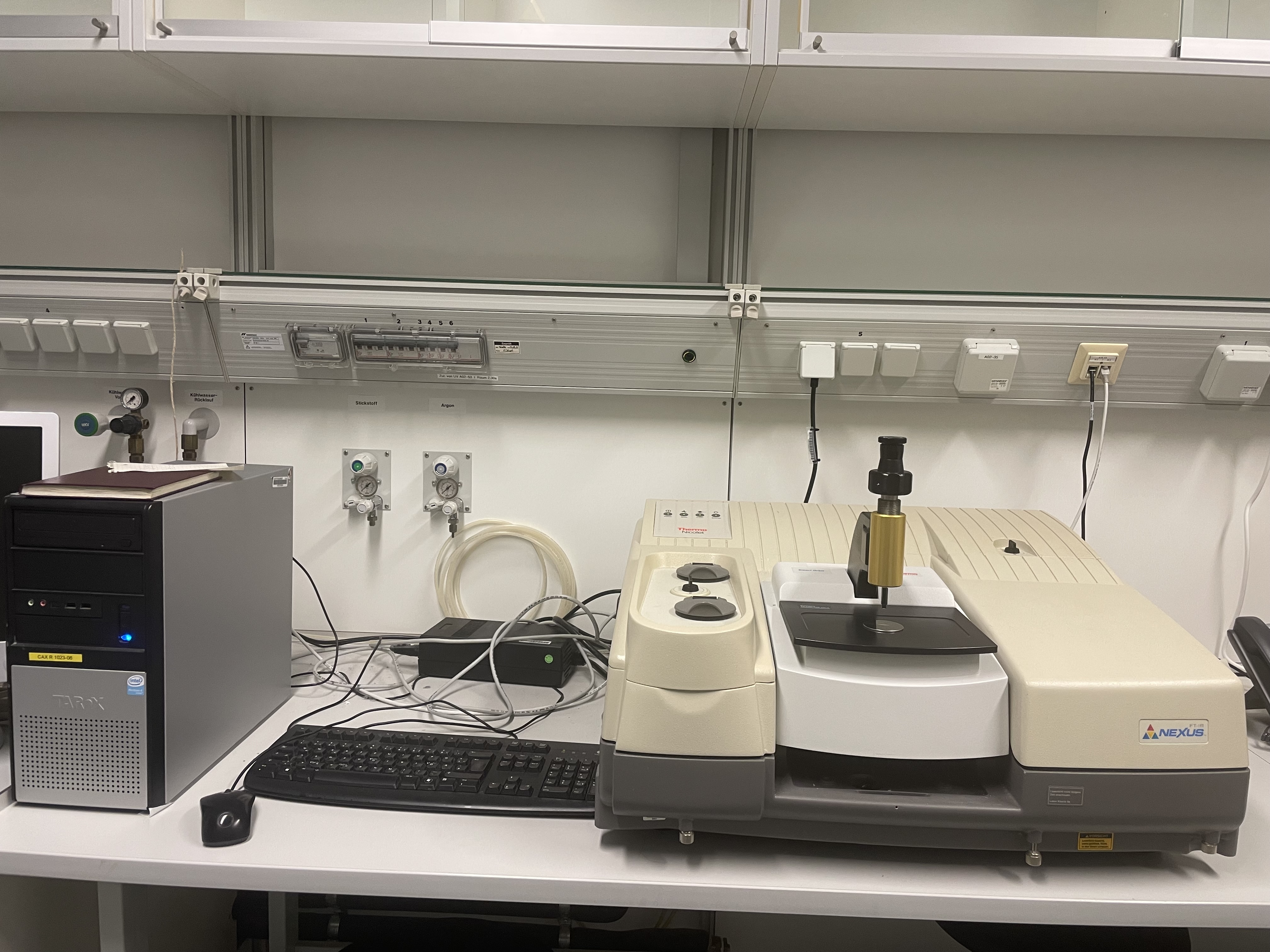
IR spectroscopy
Recording of IR spectra in transmission using KBr and CsI pellets (4000–500 cm⁻¹ and 1000–80 cm⁻¹, respectively) or by attenuated total reflection (ATR) using a diamond or ZnSe crystal (30 000–200 cm⁻¹ and 20 000–500 cm⁻¹, respectively)
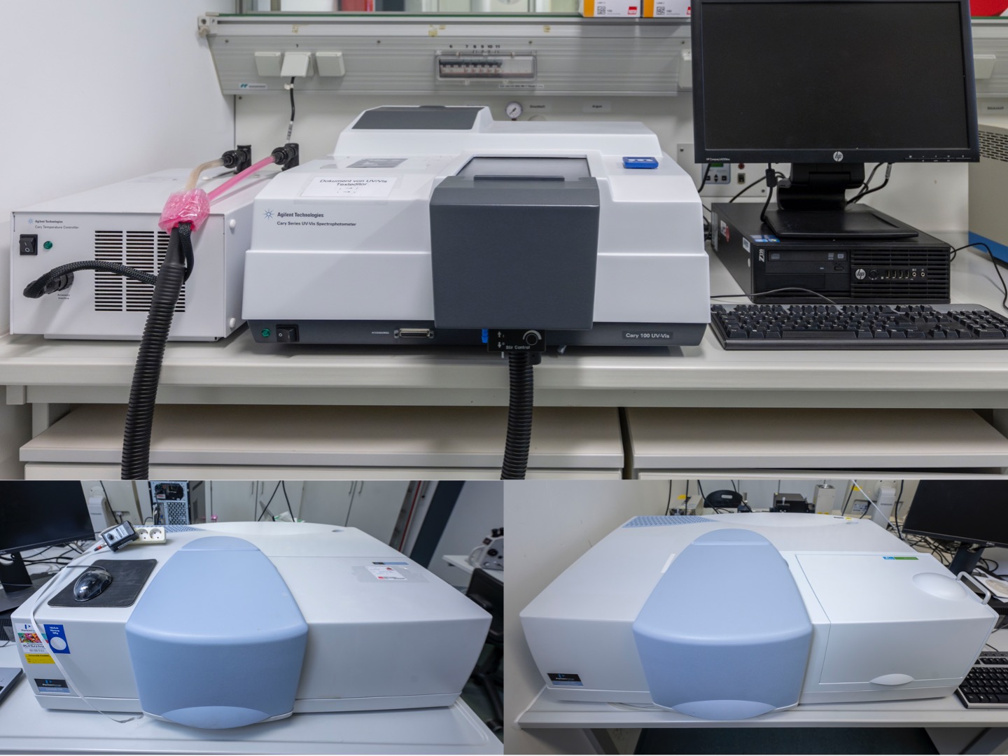
UV/VIS spectroscopy
Measurement of absorption in the 190–900 nm range (liquid and solid samples), temperature-dependent measurements with 6 cell changer
Electrochemistry
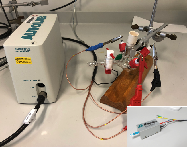
Cyclic voltammetry
Determination of redox potentials in organic solvents or aqueous solution
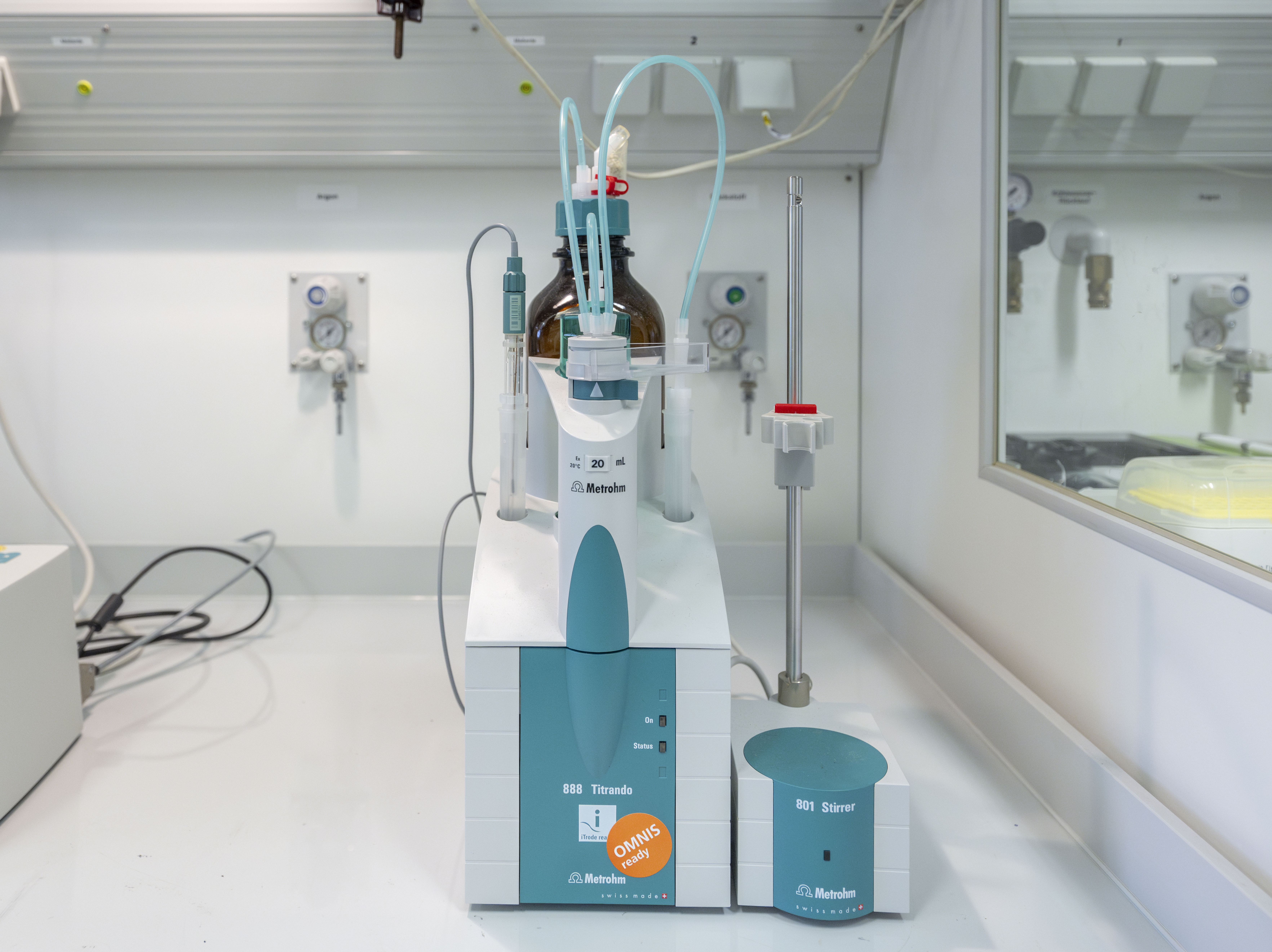
Potentiometry
Potentiometric pH titrations
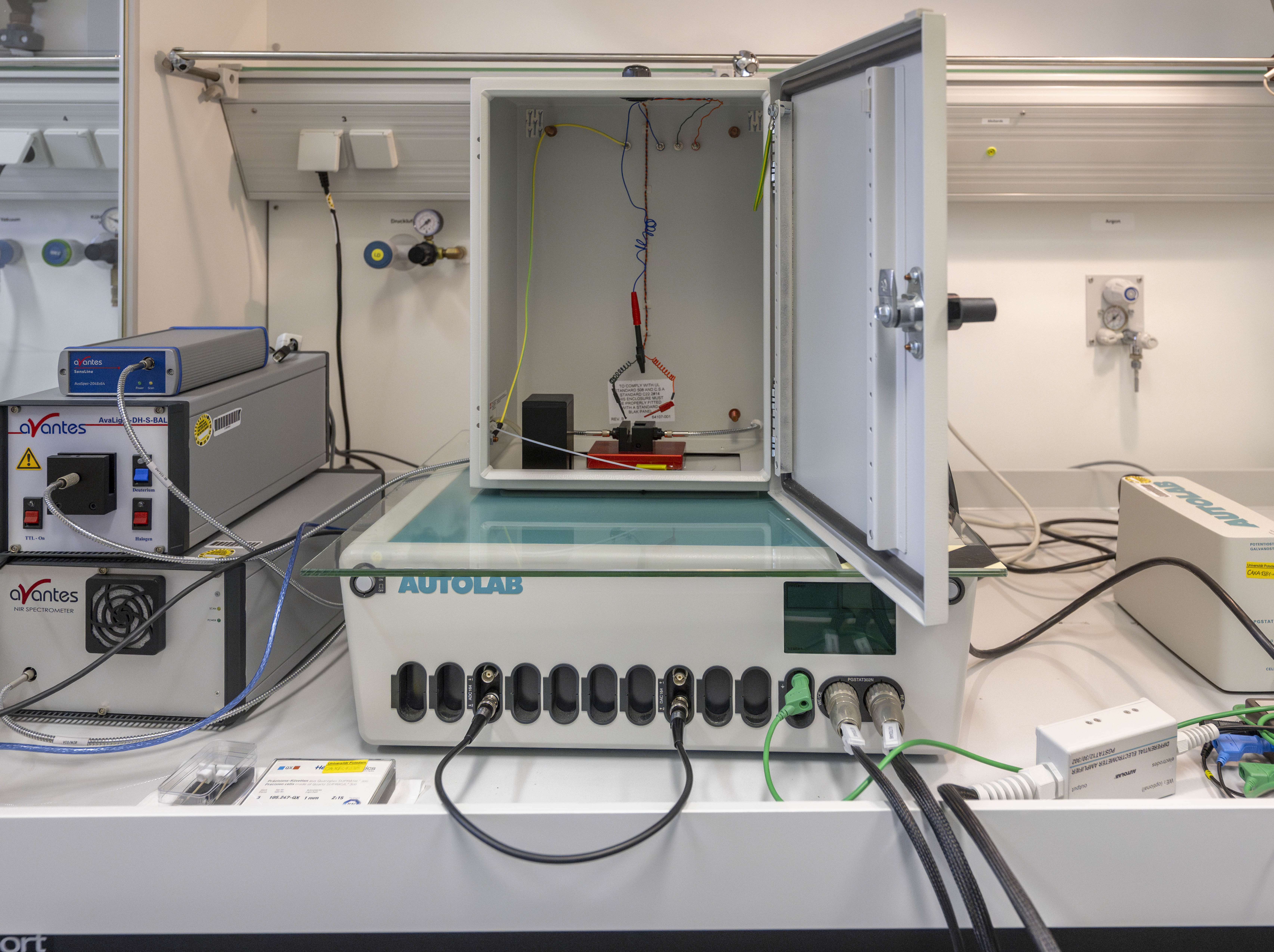
Spectroelectrochemistry
Simultaneous determination of reduction/oxidation potentials and absorption in the UV/VIS/NIR range
Element analysis
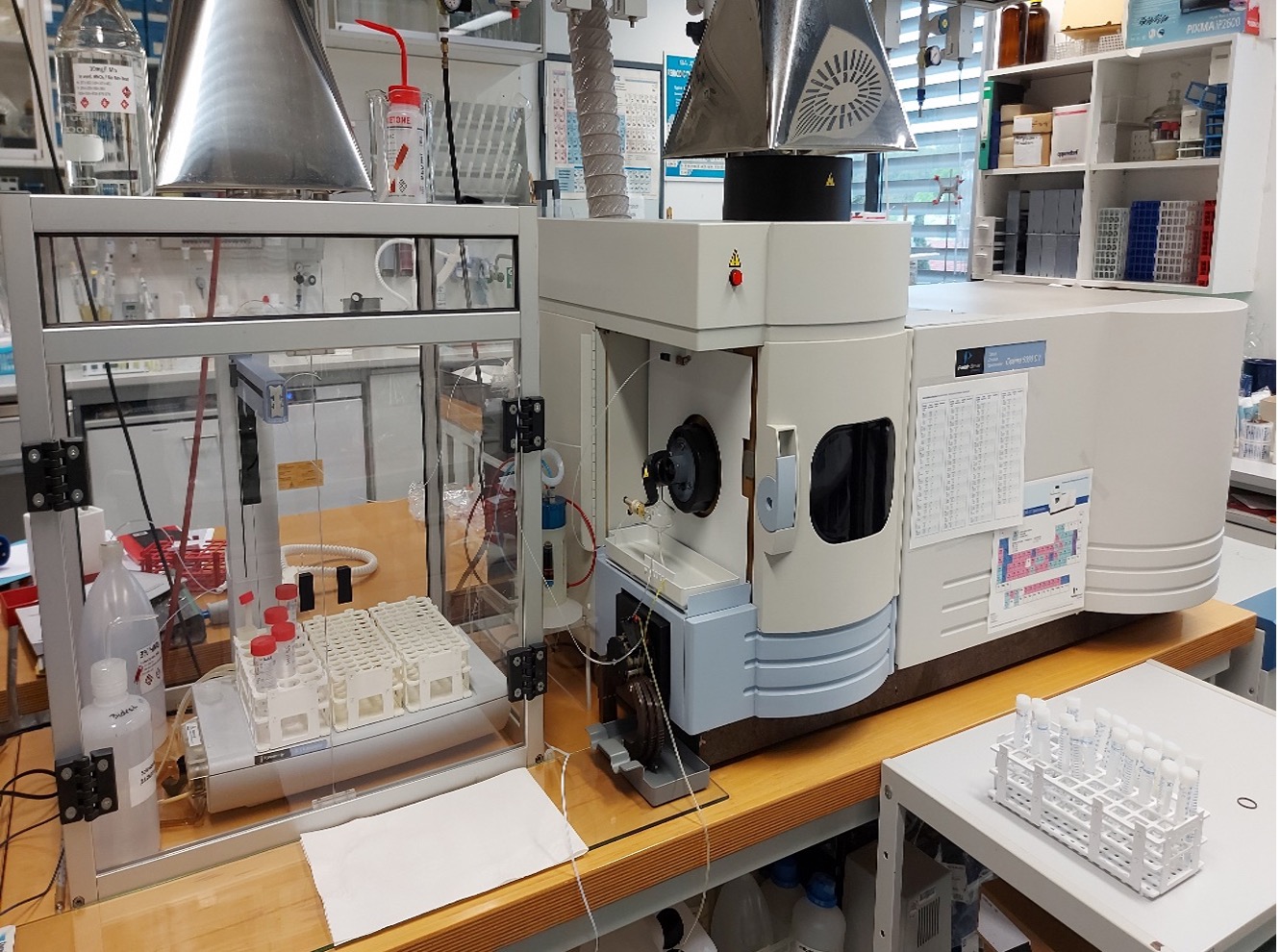
ICP-OES
Determination of elements via atomic and ionic emission (except halogens, noble gases, C, N, H, O)
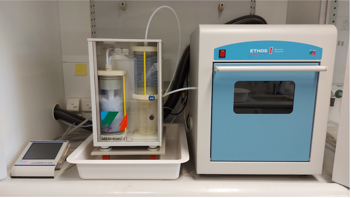
Microwave for digestion reactions
acidic and basic digestions of solid substances

Acid distillation
Purification of acids using subboiling distillation
Others
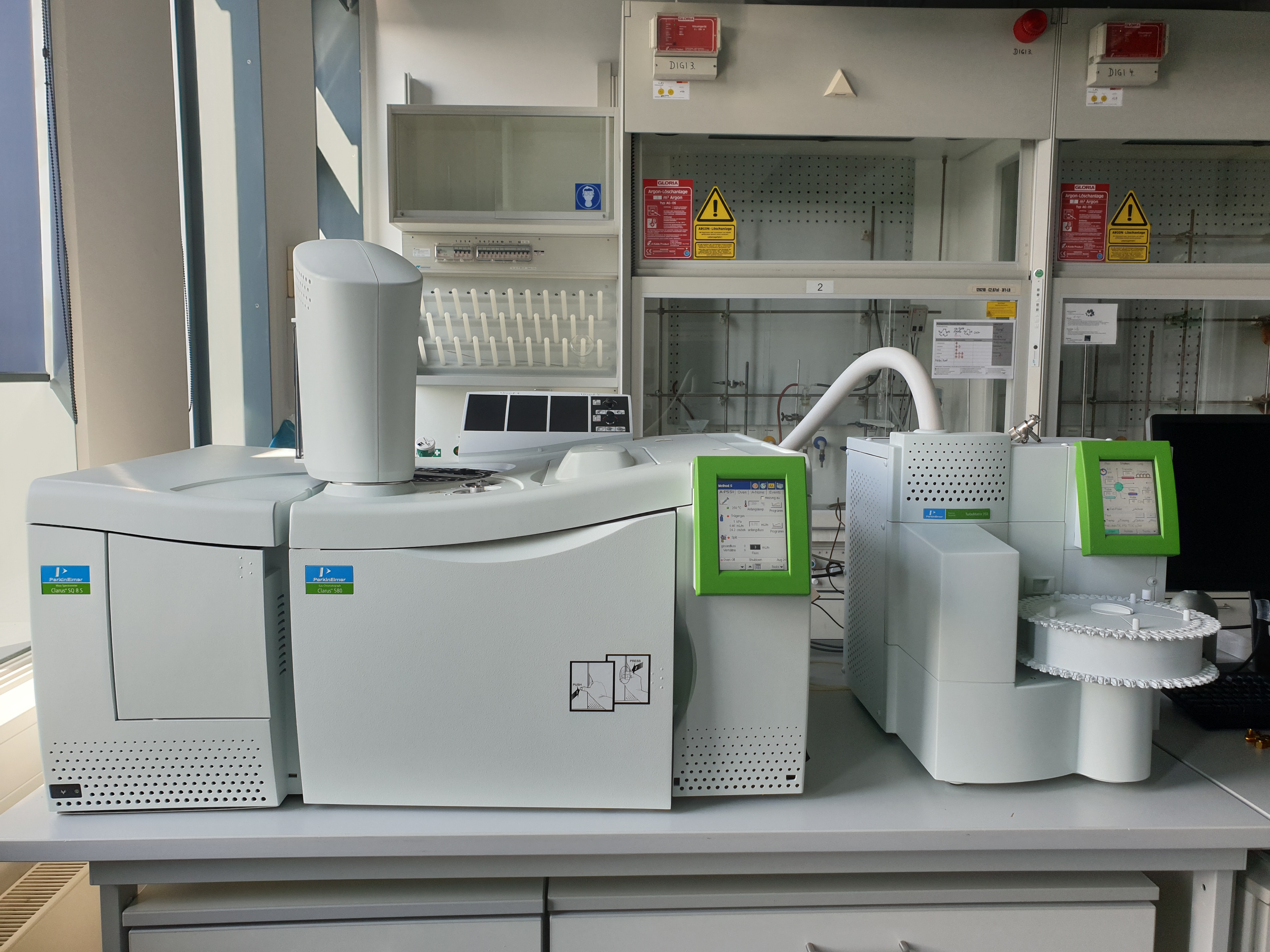
GC-MS
Separation and identification of (volatile) organic compounds by gas chromatography coupled with mass spectrometry
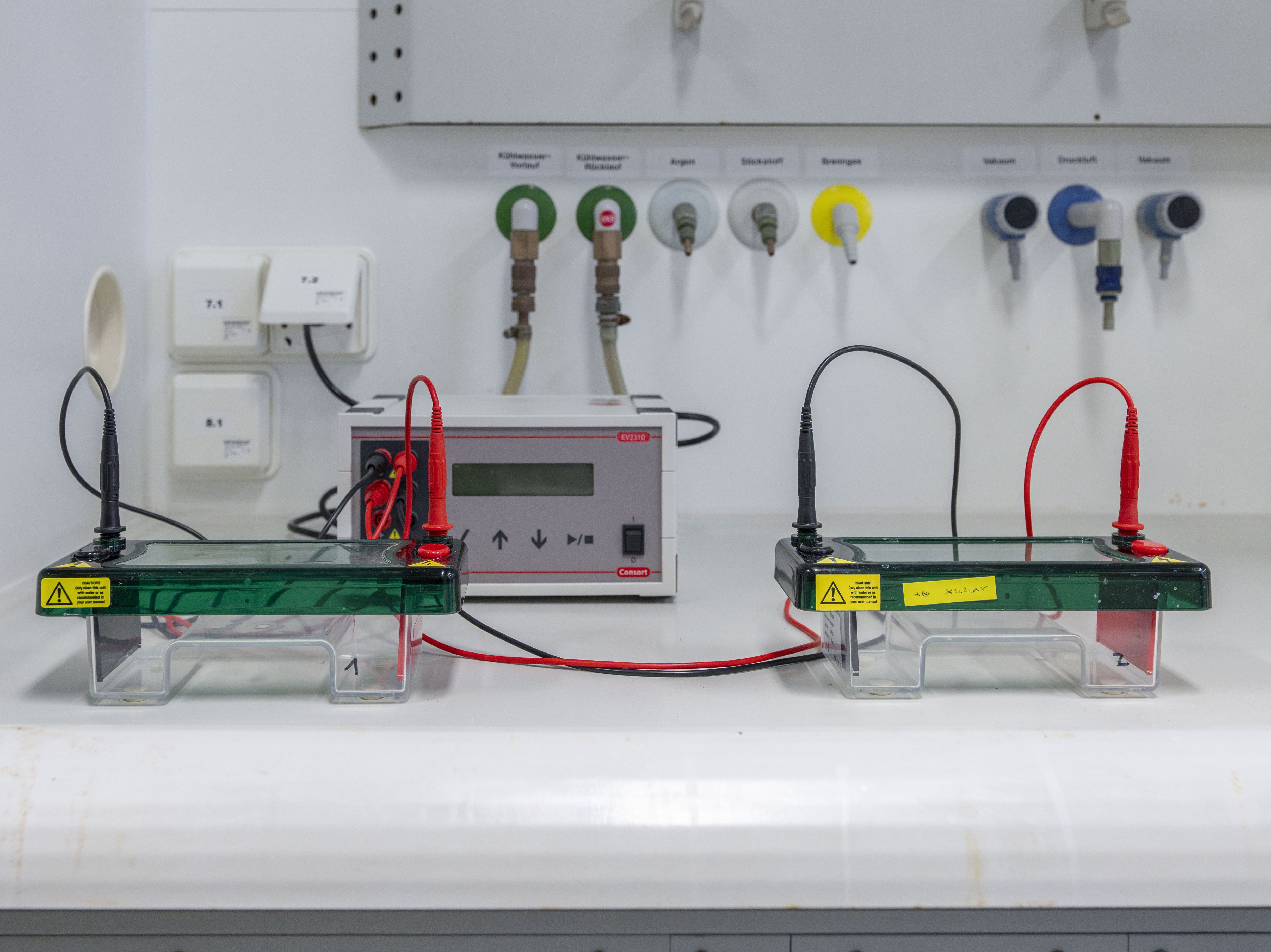
Gel electrophoresis
Separation and analysis of protein (fragments) (vertical, PAGE) and DNA (horizontal, agarose)
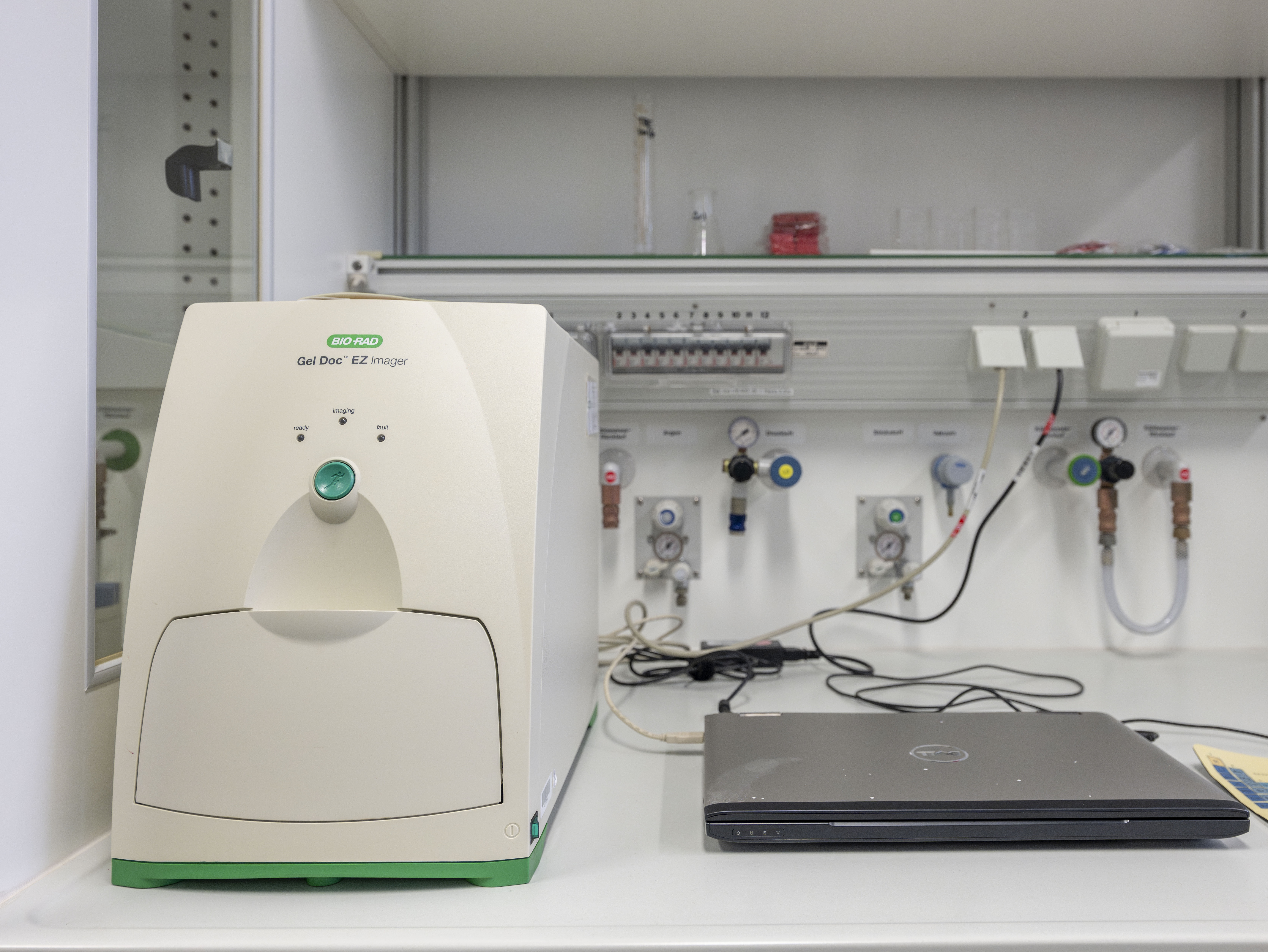
Gel scanner
Visualization und quantification of gel electrophoresis experiments with different trays depending on gel staining method
- UV Sample Tray (for irradiation with UV light)
- Stain-free Sample Tray (for stain-free gels)
- White Sample Tray (Coomassie Blue, Copper, Silver, Zinc staining)
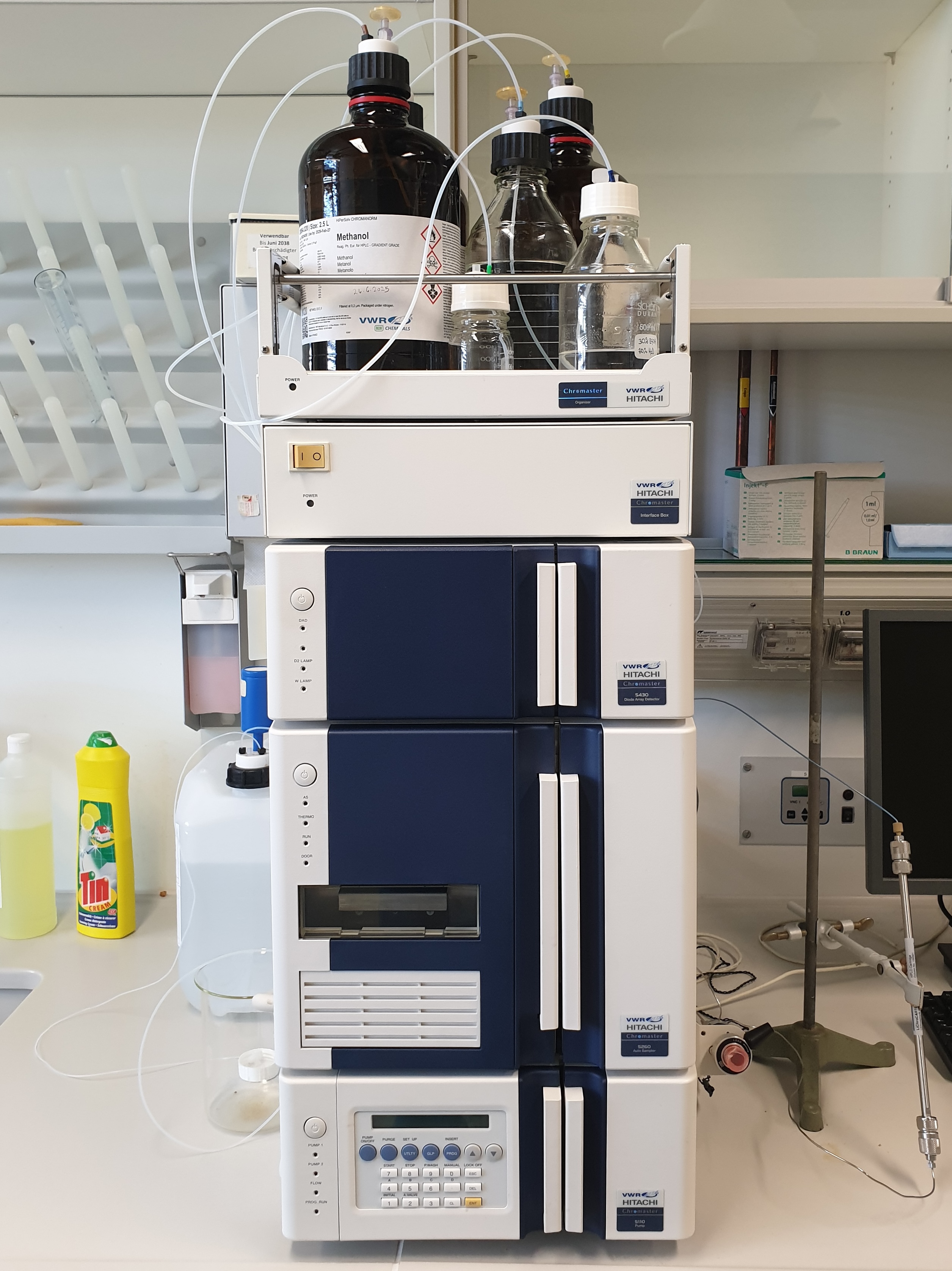
HPLC
Purification (semi-preparative, max. 50 mg) and determination of purity (analytical) with autosampler of organic compounds, especially peptides
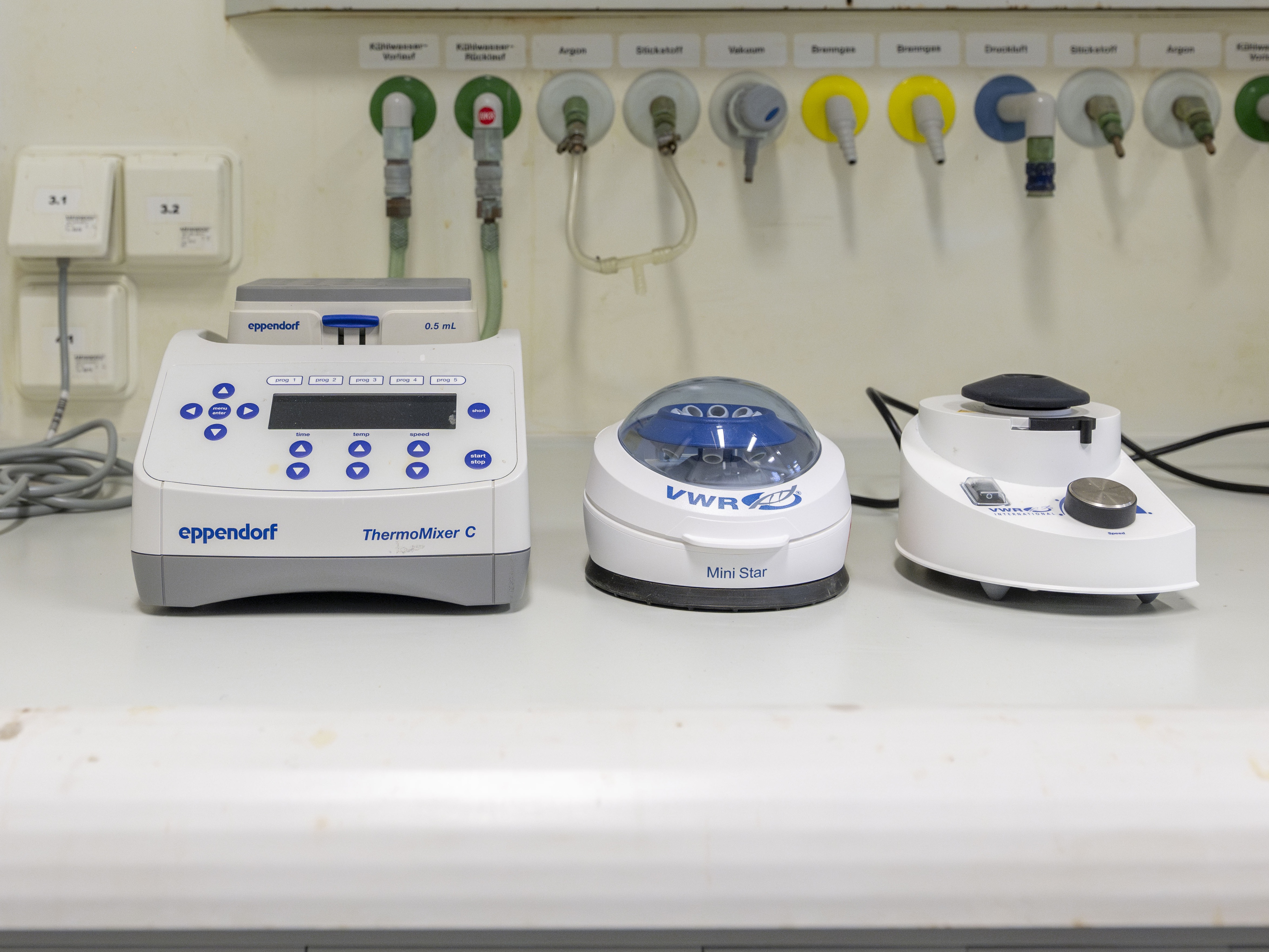
Incubation and mixing
Incubation of solution in Eppendorf tubes (0.5 and 1.5 mL) in a temperature range 1–100 °C at max. 3000 rpm, centrifugation and vortexing
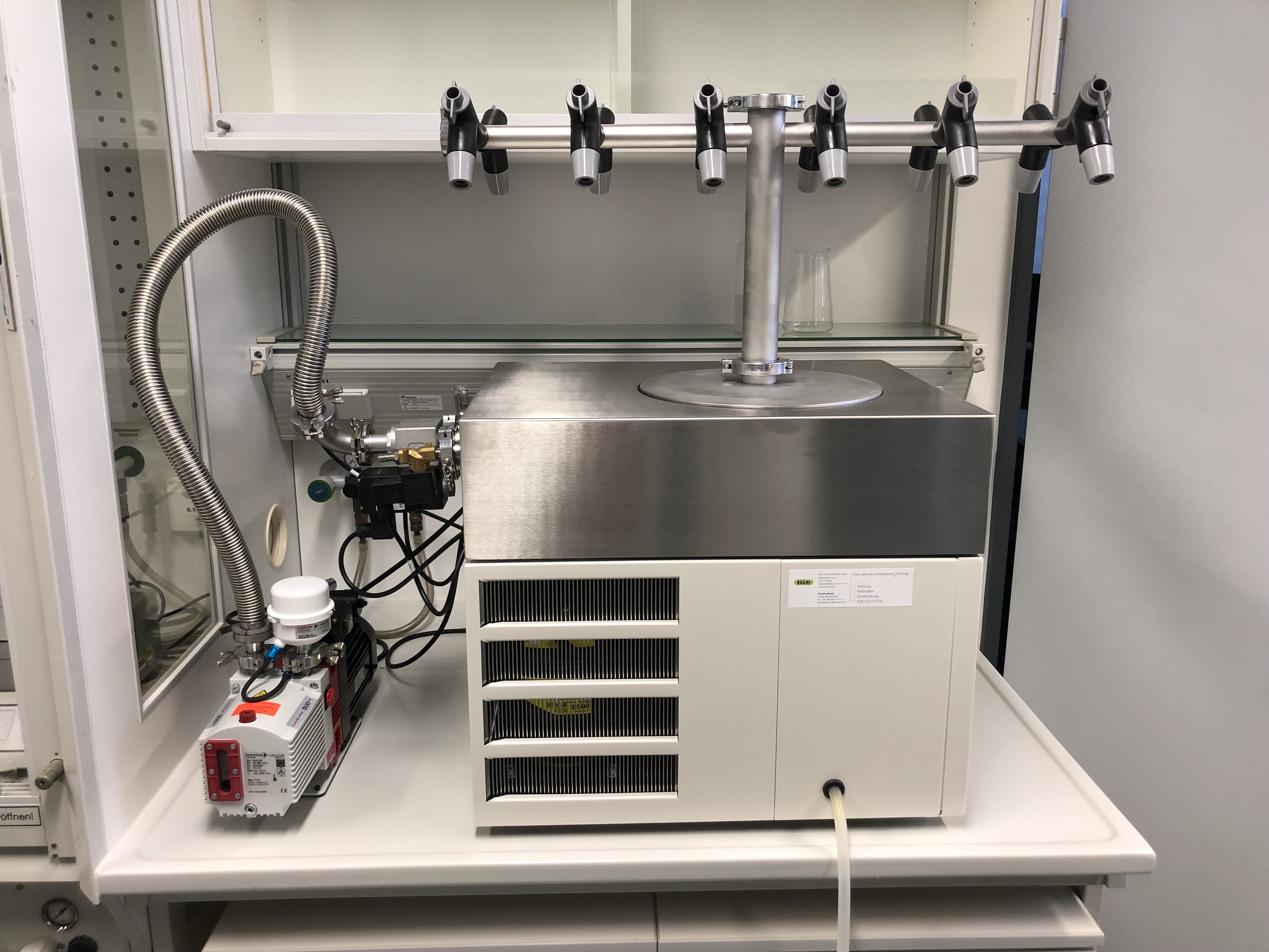
Lyophilizer
Lyophilization (freeze drying) of aqueous samples

Synthesis
- Organic synthesis and coordination chemistry, partially under inert gas atmosphere
- Reactions in parallel synthesis reactor on a small scale (irradiation possible)
- Solid phase peptide synthesis and bioconjugate chemistry

Centrifuge
Centrifugation of Falcon tubes (15 and 50 mL) and Eppendorf tubes (1,5 and 2 mL) in a temperature range of -9 to 40 °C at max. 4400 rpm