Influenza A virus (IAV) assembly at cellular membranes
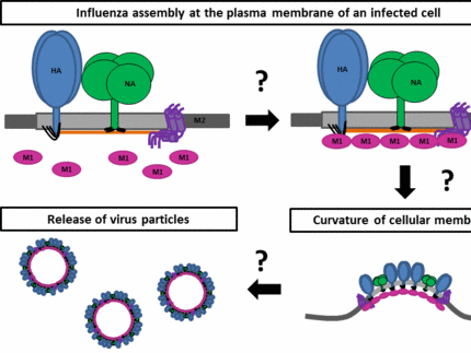
Influenza A virus (IAV) is a respiratory pathogen that causes seasonal epidemics and occasional pandemics with significant mortality. To develop innovative therapeutic approaches, a deeper understanding of the viral replication cycle is needed. One of the most abundant proteins in IAV particles is the matrix protein 1 (M1), which is essential for the virus structural stability. M1 organizes virion assembly and budding at the plasma membrane (PM), where it interacts with other viral components. The recruitment of M1 to the PM as well as its interaction with the other viral envelope proteins (hemagglutinin (HA), neuraminidase (NA), matrix protein 2 (M2)) is controversially discussed and not fully understood.
Interaction of Matrix Protein 1 with Lipid Membranes
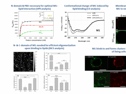
During assembly of new viral particles, M1 is recruited to the host cell membrane where it associates with lipids and other viral proteins. We employ a set of complementary experimental in vitro tools (e.g. raster image correlation spectroscopy (RICS), surface plasmon resonance (SPR) spectroscopy, circular dichroism spectroscopy, and light scattering) and in cellulo tools (e.g. number and brightness (N&B), scanning fluctuation correlation spectroscopy (sFCS)) to investigate M1-protein and M1-lipid interactions.
Recruitment of M1 to the PM and its interaction with viral envelop proteins
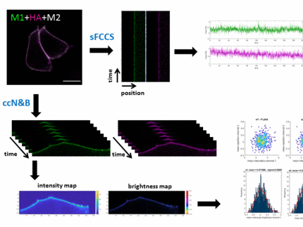
M1 is supposed to interact with viral surface proteins, but it lacks an apical transport signal, implying that the membrane localization of M1 in infected cells might be due to piggyback transport with e.g. HA, NA, and M2. For this reason, various hypotheses regarding the association of M1 to the plasma membrane (PM) have been proposed over the years. First, several studies established that M1 associates with negatively charged lipids in model membranes, but such interactions appear not to be sufficient for the actual association of M1 to the PM in non-infected cells (i.e. in cells expressing M1 as the only viral protein). Accordingly, M1 was proposed to interact with the cytoplasmic tails of HA and NA during their apical transport, as well as with the cytoplasmic tails of M2 at the assembly site. Interactions between M1 and HA, NA, or M2 have been investigated via bulk biochemistry methods (e.g. by altered detergent solubility, increased membrane association of M1 in the presence of HA or NA, or co-immunoprecipitation of M1 in the presence of M2). Nevertheless, no clear consensus has been reached regarding the role of HA, NA or M2 in recruiting M1 to the PM and its subsequent incorporation into virions. In conclusion, the molecular mechanisms involved in M1-driven IAV assembly are not fully understood and the specific interactions between M1 and other viral surface proteins have not yet been quantified directly in living cells. To obtain quantitative information on how protein-protein interactions (e.g. M1-M2 or M1-HA) occur in the native cellular environment, minimally invasive approaches (e.g. fluorescence fluctuation spectroscopy) are needed. Therefore, we apply cross-correlation Number and Brightness (ccN&B) as well as scanning fluorescence cross-correlation spectroscopy (sFCCS) analysis in living cells to quantify the oligomeric state, concentration and the diffusion dynamics of the viral envelope proteins (HA, NA, M2) and M1, as well as their interactions.
Influence of M1 mutations on virus morphology
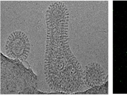
IAV virions are pleomorphic, showing morphologies that range from spherical (e.g. A/WSN/1933) to filamentous shapes (e.g. A/Udorn/301/1972). Considering this pleomorphism, it seems that virus particle morphology might play an important role in the spread and survival of the virus in nature. The filamentous morphology could assist in cell-to-cell transmission of viruses in the respiratory mucosa in vivo, whereas the spherical morphology could promote their spread through aerosols. Even though the factors determining this morphology are not completely understood, M1 has been suggested to be the key player in morphology determination, as point mutations in M1 alter virion morphology. Therefore, we want to study the effect of morphology-relevant M1 mutations from different IAV strains on M1 oligomerization behaviour and membrane binding. To address these issues, we express and purify M1 from Escherichia coli (E. coli) and its structure is analysed with circular dichroism (CD), and dynamic light scattering (DLS). Minimal membrane model systems, SLBs and GUVs, are used to analyse its binding capacity to lipids by RICS and sFCS as well as to clarify the role of M1 in membrane deformation by confocal imaging. Furthermore, we investigate the influence of these mutations on the M1 recruitment to the plasma membrane, M1 oligomerization and the interaction of M1 with other viral proteins in cellulo by using sFCCS and fluorescence recovery after photobleaching (FRAP).
Comparison of protein-protein interaction from human and avian IAV strains in human as well as avian cells
IAV can cause zoonotic infections, in which a pathogen adapted to another animal species causes disease in a human, leading to sustained human-to-human transmission and emergence of novel viruses. IAV has a multipartite RNA genome with high mutation rates. Moreover, co-infection of one host cell with two different IAVs can result in progeny viruses containing gene segments of both parental viruses. Both factors can facilitate such host adaptions. The genetic contribution of the M segment (encoding M1 and M2) to host adaptation and the underlying mechanisms are not yet understood. A comparison of IAVs with avian- and human-derived M segments in avian and mammalian systems revealed that the avian M segment restricts viral growth specifically in mammalian cells. It is not yet clear to what extent the co-expression of viral proteins with different host origins affects protein-protein interaction (M1-M1 and M1-M2) and the recruitment of M1 to the plasma membrane. We investigate this topic using quantitative fluorescence microscopy methods and human/avian cell models.
Funding:
Our research project is funded by the German Research Foundation.


