Spectroscopy
Fluorescence spectroscopy
In our lab we work with standard fluorescence measurements and we are using several fluorescence based techniques like FCS and FRET. For instance we observe protein folding and carbohydrate binding using the intrinsic protein fluorescence. By fluorescently labeling carbohydrates we determine hydrolysis rates of carbohydrate specific enzymes. We monitor in vitro DNA-ejection of bacteriophages in presence of a DNA-staining dye in fluorescence.
We have several fluorescence spectrometers from Jasco and Horiba.
Circular dichroism (CD) spectroscopy
As optically active molecules, proteins give rise to CD absorption bands in the far and near UV spectral range. We are using CD spectroscopy to monitor protein folding and stability on the secondary and tertiary structure level. This gives us information on overall protein conformation in aqueous solution and in solutions of increasing osmolality.
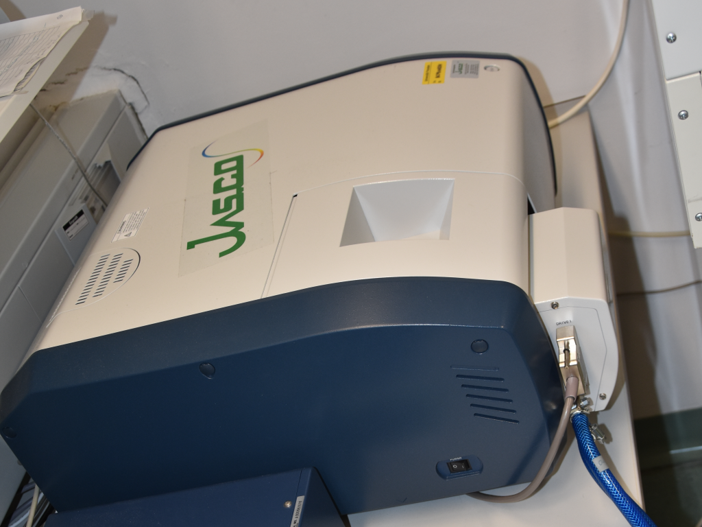
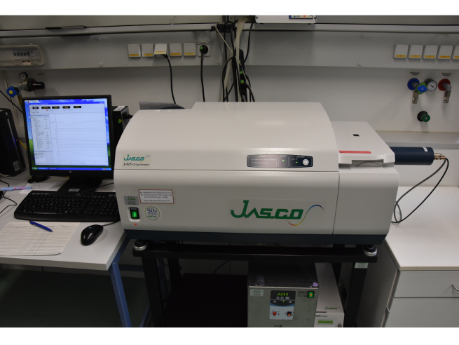
We have two Jasco CD spectropolarimeters, a J-815 and a J-715.
Reichert SR7500DC Zweikanal SPR Biosensor
Im Rahmen einer EFRE-unterstützten Infrastrukturmaßnahme wurde die Finanzierung eines Spektroskops zur Messung von Oberflächenplasmonresonanz (surface plasmon resonance, SPR) gefördert. Das Gerät wird für Forschungs- und Entwicklungsvorhaben mit Schwerpunkt in den Arbeitsgruppen Physikalische Biochemie und Molekulare Biotechnologie an der Universität Potsdam erfolgreich eingesetzt. Die Arbeitsgruppe Physikalische Biochemie konnte damit einen Sensor für bakterielle Oberflächenkohlenhydrate entwickeln. Das optisch-diagnostische Verfahren detektiert Pathogene ohne zeitaufwändige Zellkulturexperimente schnell und mit sehr hoher Sensitivität. Das Detektionssystem basiert auf einer stabilen Proteinkomponente aus Bakteriophagen, die die bakterielle Zellwand erkennt. Dieses Verfahren wurde zum Patent angemeldet. Durch das mit EFRE-Unterstützung beschaffte Gerät konnten wir Oberflächen-basierte Screening-Verfahren entwickeln, mit denen die Sensitivität der Protein-Bakterien-Interaktionen quantifiziert werden kann und die Sensorkomponenten auf Spezifität und Affinität getestet werden können. Wir haben damit z.B. Lipopolysaccharid- Zellwandpräparationen des Darmpathogens Shigella flexneri immobilisiert und die Interaktion mit den Sensorproteinen aus Bakteriophagen gemessen.
